3rd Degree AV Block on the ECG
Third-degree atrioventricular block is complete failure of conduction between atria and ventricles, in which P waves and QRS complexes occur independently at their own rates and an escape rhythm maintains ventricular activity.
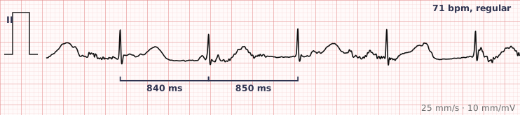
ECG criteria
| Feature | What you see in 3rd Degree AV Block |
|---|---|
| Conduction | None. No P wave conducts. Atria and ventricles are electrically independent. |
| P waves | Regular, at the sinus rate — usually faster than the QRS rate. Some fall on QRS complexes and T waves. |
| QRS rate | Slower and regular, set by the escape focus: 40–60 if junctional, 20–40 if ventricular. |
| PR interval | Varies randomly, because there is no relationship to measure. This is the diagnostic feature. |
| QRS width | Narrow if the escape is junctional; wide if it is ventricular. Narrower and faster is more stable. |
| Rhythm | Both the P waves and the QRS complexes are individually regular, but unrelated to each other. |
How to spot it
- Notice a slow, regular ventricular rhythm — that combination should always prompt a search for dissociation.
- March out the P waves across the whole strip. They should be regular at their own rate, and there will be more of them than QRS complexes.
- March out the QRS complexes separately. They should also be regular, at a slower rate.
- Measure several PR intervals. If they vary randomly, the two are dissociated — that is complete heart block.
- Hunt for P waves hidden inside T waves and QRS complexes. Missing them is the usual reason the diagnosis is delayed.
- Assess the escape: narrow and 40–60 is junctional and relatively stable; wide and 20–40 is ventricular and unstable.
What it gets confused with
| Looks like | How to tell them apart |
|---|---|
| Sinus bradycardia | Every QRS follows a P wave at a fixed PR. In complete block the relationship is random. |
| 2:1 or high-grade second-degree block | Some P waves still conduct with a consistent PR. In complete block none do. |
| Junctional escape rhythm | The escape rhythm is the consequence of the block, not an alternative diagnosis — but without visible independent P waves it can look like isolated junctional rhythm. |
| Ventricular tachycardia with AV dissociation | Also dissociated, but fast. Complete heart block is slow. |
| Atrial fibrillation with complete block | No P waves at all; a regular slow ventricular rate in known AF. Classically digoxin toxicity. |
Traps
- The diagnosis is made by proving the PR interval varies, not by counting rates. Measure several PR intervals deliberately before committing.
- P waves buried in QRS complexes and T waves are constantly missed. Use calipers, march the atrial rate out, and mark where each P must fall.
- Do not suppress the escape rhythm. It is the only thing generating cardiac output.
- Complete heart block with a wide, slow escape is unstable and can deteriorate to asystole. Treat it as an emergency requiring pacing.
- New complete heart block in the context of an inferior myocardial infarction is often transient and vagally mediated; in an anterior infarction it signals extensive septal damage and carries a much worse prognosis.
Why it happens
Conduction fails completely at the AV node, the His bundle, or both bundle branches. The atria continue under sinus control while the ventricles are driven by whichever subsidiary pacemaker below the block has the fastest intrinsic rate. Because the two pacemakers are entirely independent, the timing relationship between P waves and QRS complexes is random.
Why it matters
Complete heart block is a medical emergency when the escape rhythm is slow or wide, because such escapes are unreliable and may stop. It is the classic indication for pacing — temporary if there is a reversible cause such as drug toxicity or inferior infarction, permanent if the conduction system is irreversibly diseased.
Questions
How do I recognise complete heart block on an ECG?
Find a slow regular ventricular rhythm, then march out the P waves separately. If P waves are regular at their own faster rate and the PR interval varies randomly from beat to beat, the atria and ventricles are dissociated and the block is complete.
What is AV dissociation?
Atria and ventricles beating independently, each driven by its own pacemaker. It is the defining feature of complete heart block, and it also occurs in ventricular tachycardia — the difference being that complete heart block is slow and VT is fast.
Why does the escape rhythm matter?
It tells you where the block is and how stable the patient is. A narrow escape at 40 to 60 arises in the AV junction and is comparatively reliable. A wide escape at 20 to 40 arises in ventricular tissue, is much less reliable, and can fail altogether.
Is complete heart block always permanent?
No. It can be reversible when caused by drugs, electrolyte disturbance, or an inferior myocardial infarction where the block is often vagally mediated and temporary. Block complicating an anterior infarction usually reflects extensive irreversible damage.
Reading about 3rd degree av block is not the same as calling it on a tracing you have never seen.
Practise on real cases in ECG Pro