Atrial Fibrillation (AF) on the ECG
Atrial fibrillation is a supraventricular arrhythmia in which the atria depolarise chaotically, producing an ECG with no discernible P waves and an irregularly irregular ventricular response.
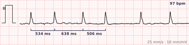
ECG criteria
| Feature | What you see in AF |
|---|---|
| Rate | Ventricular rate varies widely. Untreated it is often 100–180; on rate control it may be 60–100. |
| Rhythm | Irregularly irregular — no pattern to the R-R intervals at all. This is the single most reliable sign. |
| P wave | Absent. Replaced by fibrillatory waves, which may be coarse (over 0.5 mm) or so fine the baseline just looks noisy. |
| PR interval | Not measurable — there is no P wave to measure from. |
| QRS | Narrow, under 120 ms, unless there is bundle branch block, an accessory pathway, or rate-related aberrancy. |
| ST-T | Often non-specific. Rate-related ST depression is common and does not by itself mean ischaemia. |
How to spot it
- Take a caliper or a piece of paper to the rhythm strip and mark three consecutive R waves. Move it along. In AF the marks never line up again — that is irregular irregularity.
- Look for P waves in lead II and V1, the two leads where atrial activity shows best. In AF there are none.
- Check the baseline between QRS complexes. Coarse AF has visible undulating fibrillatory waves; fine AF has a baseline that simply never sits still.
- Confirm the QRS is narrow. A wide, irregular, fast rhythm raises AF with pre-excitation, which is a different and more dangerous problem.
- Report the ventricular rate as a range rather than a single number, because in AF it genuinely varies from beat to beat.
What it gets confused with
| Looks like | How to tell them apart |
|---|---|
| Atrial flutter with variable block | Flutter has organised sawtooth waves at about 300/min. AF has no organised atrial activity. Look hard at V1. |
| Multifocal atrial tachycardia | Irregular, but P waves are present with at least three different shapes. |
| Frequent premature atrial contractions | An underlying regular sinus rhythm interrupted by early beats — the underlying rhythm is still findable. |
| Sinus arrhythmia | P waves present and normal; the variation is gradual and follows respiration. |
| AF with rapid ventricular response | The same rhythm conducting fast — over 100, often 110–180. Still narrow, still irregular. |
| AF with pre-excitation | Irregular, very fast (often over 200) and wide, with varying QRS width. A dangerous variant to recognise. |
Traps
- At very fast rates AF starts to look regular. As the rate climbs, the absolute variation between R-R intervals shrinks and the eye stops seeing it. Measure rather than judge by appearance.
- Fine fibrillatory waves are easily read as artefact or as P waves. Baseline wander and tremor both mimic them — check whether the "P waves" bear any consistent relationship to the QRS complexes. In AF they do not.
- A regular ventricular response in known AF is not reassuring: it suggests complete heart block with a junctional escape, classically from digoxin toxicity.
- Never give AV nodal blocking drugs to irregular, wide, fast AF. If an accessory pathway is conducting, blocking the AV node pushes more impulses down it.
Why it happens
Disorganised re-entrant wavelets in the atria, commonly triggered by ectopic foci around the pulmonary vein ostia, replace coordinated atrial depolarisation. The atria quiver rather than contract, so there is no P wave, and the AV node is bombarded at 400–600 impulses per minute. It conducts only some of them, and unpredictably, which is what makes the ventricular response irregular.
Why it matters
Atrial fibrillation is the most common sustained arrhythmia, and its importance is out of proportion to how it feels. Loss of atrial contraction costs perhaps 20% of cardiac output, and stasis in the left atrial appendage raises stroke risk roughly fivefold. Recognising it on a tracing frequently matters more for the stroke risk it reveals than for the symptoms it causes.
Questions
What is the single most reliable sign of atrial fibrillation on an ECG?
An irregularly irregular ventricular rhythm with no P waves. Absent P waves alone are not enough, since junctional rhythms also lack them; it is the combination with genuine irregularity that makes the diagnosis.
Can atrial fibrillation ever look regular?
Yes, in two situations. At very fast rates the beat-to-beat variation becomes hard to see by eye. And if complete heart block develops, an escape rhythm takes over and becomes regular — classically a sign of digoxin toxicity.
What is the difference between coarse and fine atrial fibrillation?
Only the amplitude of the fibrillatory waves — coarse is above about 0.5 mm, fine below. The distinction has little clinical consequence; fine AF is simply harder to distinguish from a noisy baseline.
Why is a wide, fast, irregular rhythm dangerous?
It suggests atrial fibrillation conducting down an accessory pathway rather than through the AV node. Rates can be extremely high and the rhythm can degenerate into ventricular fibrillation. AV nodal blocking drugs make it worse.
Reading about atrial fibrillation is not the same as calling it on a tracing you have never seen.
Practise on real cases in ECG Pro