Wandering Atrial Pacemaker (WAP) on the ECG
Wandering atrial pacemaker is an irregular supraventricular rhythm at a rate below 100 in which the pacemaking site shifts between the sinus node and other atrial foci, producing at least three distinct P wave morphologies in a single lead.
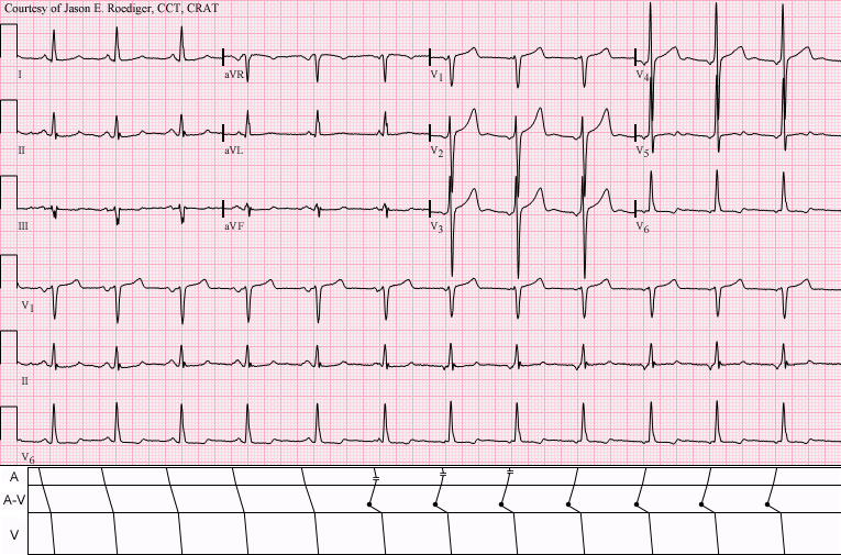
ECG criteria
| Feature | What you see in WAP |
|---|---|
| Rate | Under 100 bpm. The same picture above 100 is called multifocal atrial tachycardia. |
| Rhythm | Irregular, because each focus fires at its own intrinsic rate. |
| P wave | At least three different shapes in one lead. Every QRS still has a P wave in front of it. |
| PR interval | Varies with the P wave shape — a focus closer to the AV node gives a shorter PR. |
| QRS | Narrow, under 120 ms, and constant in shape. |
| ST-T | Usually normal. |
How to spot it
- Pick one lead with clear P waves — lead II or V1 — and stay in it. Comparing P shapes across different leads proves nothing.
- Work along the strip and group the P waves by shape. Three or more distinct morphologies makes the diagnosis.
- Confirm each QRS still has a P in front of it. That is what separates this from atrial fibrillation.
- Measure a few PR intervals. They should vary in step with the P wave shape.
- Check the rate. Under 100 it is wandering atrial pacemaker; over 100 the same finding is multifocal atrial tachycardia.
What it gets confused with
| Looks like | How to tell them apart |
|---|---|
| Atrial fibrillation | No P waves at all. WAP has plenty of P waves, just differently shaped ones. |
| Frequent premature atrial contractions | An underlying regular sinus rhythm with intermittent early beats, rather than a continuously shifting pacemaker. |
| Multifocal atrial tachycardia | The same rhythm at a rate above 100. Strongly associated with severe lung disease. |
| Sinus arrhythmia | P waves all identical; only the rate varies, and it varies with breathing. |
Traps
- Count P wave shapes in a single lead only. The same P wave looks different in different leads by definition, so comparing across leads generates false morphologies.
- Wandering atrial pacemaker is frequently misread as atrial fibrillation because both are irregular. The presence of P waves is the whole distinction.
- It is often a normal variant, particularly in the young and in athletes with high vagal tone, and needs no investigation on its own.
- When the same pattern runs above 100, look hard for underlying COPD, hypoxia or theophylline — multifocal atrial tachycardia is rarely idiopathic.
Why it happens
Dominance passes back and forth between the sinus node and one or more ectopic atrial foci, usually because vagal tone suppresses the sinus node enough to let slower foci escape. Each focus depolarises the atria from a different starting point, so each produces a P wave of different shape and axis, and a different conduction time to the AV node.
Why it matters
In isolation and at a normal rate, wandering atrial pacemaker is usually benign and needs nothing. Its value is mainly as a distinguishing skill: recognising P waves in an irregular rhythm prevents a mistaken diagnosis of atrial fibrillation and the anticoagulation decision that follows from it.
Questions
How many P wave shapes are needed for the diagnosis?
At least three distinct morphologies, counted within a single lead. Two shapes is usually just sinus rhythm with an ectopic focus.
What is the difference between wandering atrial pacemaker and multifocal atrial tachycardia?
Only the rate. The same multiple-P-wave picture is called wandering atrial pacemaker below 100 beats per minute and multifocal atrial tachycardia above it. The faster form is strongly associated with severe lung disease.
Is wandering atrial pacemaker dangerous?
On its own, usually not. It is common in young people and athletes with high vagal tone and often needs no treatment or investigation.
Reading about wandering atrial pacemaker is not the same as calling it on a tracing you have never seen.
Practise on real cases in ECG Pro