Escape Rhythm on the ECG
An escape rhythm is a slow rhythm generated by a subsidiary pacemaker in the AV junction or ventricle when the sinus node fails or its impulses are not conducted, with a rate and QRS width that reveal how far down the conduction system the escape focus lies.
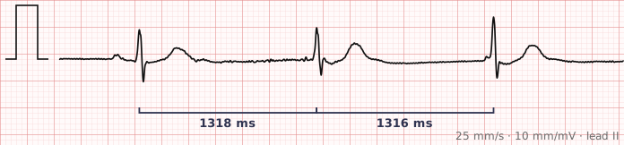
ECG criteria
| Feature | What you see in Escape Rhythm |
|---|---|
| Rate | Junctional 40–60. Ventricular 20–40. The lower the focus, the slower the rate. |
| Rhythm | Regular, because a single automatic focus is driving it. |
| P wave | Absent, inverted, or falling after the QRS in junctional escape. Dissociated in ventricular escape. |
| PR interval | Short or absent if a retrograde P precedes the QRS; otherwise not measurable. |
| QRS | Narrow in junctional escape (the His-Purkinje system is used normally). Wide in ventricular escape, over 120 ms. |
| ST-T | Discordant to the QRS in ventricular escape, as with any wide complex. |
How to spot it
- Note that the rate is slow and the rhythm regular — that combination should immediately raise escape.
- Measure the QRS width. Narrow points to a junctional focus; wide points to a ventricular one.
- Use the rate to confirm: 40–60 fits junctional, 20–40 fits ventricular. The two measurements should agree.
- Look for P waves. Absent, inverted or retrograde P waves fit junctional escape; P waves marching independently fit ventricular escape with complete block above.
- Then ask what the escape is compensating for — a failing sinus node, or complete heart block. The escape rhythm is the consequence, not the diagnosis.
What it gets confused with
| Looks like | How to tell them apart |
|---|---|
| Sinus bradycardia | Normal upright P wave before every QRS with a constant PR. Escape rhythms have no such P wave. |
| Complete heart block | Not an alternative so much as the usual cause. Independent P waves at a faster rate confirm it. |
| Accelerated idioventricular rhythm | The same wide escape morphology running at 50–110, classically after reperfusion of an infarct. |
| Ventricular tachycardia | Wide, like ventricular escape, but fast. Rate alone separates them. |
| Paced rhythm | Pacing spikes before each complex. |
Traps
- Never suppress an escape rhythm. It is the only thing keeping the patient alive. Drugs given to "treat" what looks like an ectopic focus can produce asystole.
- A slow wide-complex rhythm is easy to mistake for a ventricular arrhythmia. Look at the rate: 20–40 is escape, not tachycardia.
- The escape rate tells you where the block is. A narrow escape at 50 is reassuring; a wide escape at 25 is unstable and unlikely to be sustained.
- Always look for what the escape is escaping from — the important diagnosis is usually in the P waves, not the QRS complexes.
Why it happens
Cardiac tissue below the sinus node has its own intrinsic automaticity, but is normally overdriven and suppressed by faster impulses from above. When those impulses stop arriving, the fastest remaining focus takes over. The AV junction depolarises at 40–60 and reaches the ventricles through the normal His-Purkinje system, so the QRS is narrow; a ventricular focus depolarises at 20–40 and spreads slowly through muscle, so the QRS is wide.
Why it matters
An escape rhythm is a safety mechanism doing its job, and its characteristics are a direct readout of how far down the conduction system has failed. A narrow junctional escape at 50 is comparatively stable. A wide ventricular escape at 25 signals failure low in the conduction system, is prone to stopping altogether, and usually means pacing.
Questions
Why is a junctional escape rhythm narrow and a ventricular one wide?
A junctional focus sits above the bundle branches, so the impulse still travels through the fast His-Purkinje system and depolarises both ventricles quickly. A ventricular focus is below that system, so the impulse spreads slowly through muscle, taking longer and producing a wide QRS.
What rate should an escape rhythm be?
It depends on the focus. The AV junction fires intrinsically at 40 to 60 beats per minute, and ventricular tissue at 20 to 40. The rate is therefore a good guide to how far down the escape has originated.
Should an escape rhythm ever be suppressed?
No. An escape rhythm exists because the pacemaker above it has failed, and it is maintaining cardiac output. Suppressing it can cause asystole. The correct approach is to treat the underlying cause of the failure.
Reading about escape rhythm is not the same as calling it on a tracing you have never seen.
Practise on real cases in ECG Pro