Pacemaker Rhythm on the ECG
A pacemaker rhythm is a cardiac rhythm generated by an implanted pacing device, identified on the ECG by sharp pacing spikes immediately preceding the chamber they capture, with a paced QRS morphology determined by the position of the pacing lead.
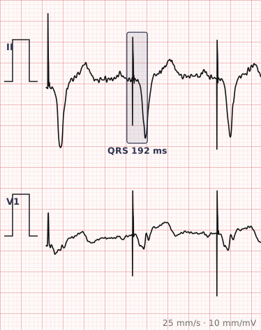
ECG criteria
| Feature | What you see in Pacemaker Rhythm |
|---|---|
| Pacing spike | A sharp, narrow vertical deflection immediately before the captured complex. May be very small on digital recordings. |
| Atrial pacing | Spike followed by a P wave. QRS is normal if conduction below is intact. |
| Ventricular pacing | Spike followed by a wide QRS. Right ventricular apical pacing gives an LBBB-like pattern with left axis deviation. |
| Dual chamber | Two spikes per cycle — one before the P wave, one before the QRS. |
| ST-T | Discordant to the paced QRS, as with any wide complex. |
| Rate | At or above the programmed lower rate limit. Sudden rate changes may be normal device behaviour. |
How to spot it
- Look for pacing spikes. On modern digital recordings they can be tiny — scan every lead, since a spike perpendicular to one lead may be invisible in it.
- Work out which chamber is paced by seeing what follows each spike: a P wave means atrial, a wide QRS means ventricular, both means dual chamber.
- Check for capture — every spike should be followed by a depolarisation. A spike with nothing after it is failure to capture.
- Check for appropriate sensing. Spikes falling on top of intrinsic beats suggest undersensing; unexpectedly long pauses suggest oversensing.
- Note the paced QRS morphology. Right ventricular pacing normally looks like LBBB; a right bundle branch block pattern raises the possibility of lead displacement.
- Do not attempt to read ischaemia from the paced complexes directly — the repolarisation is already abnormal.
What it gets confused with
| Looks like | How to tell them apart |
|---|---|
| Left bundle branch block | Same broad morphology but no pacing spikes. Look carefully before deciding. |
| Ventricular tachycardia | Wide and fast with no spikes. A malfunctioning device can also drive a fast paced rhythm. |
| Escape rhythm | Wide and slow with no spikes — the natural equivalent of what pacing replaces. |
| Fusion and pseudofusion beats | Intrinsic conduction and pacing coinciding. Normal device behaviour, not malfunction. |
| Artefact | Spike-like deflections not followed by any consistent depolarisation and not at a regular interval. |
Traps
- Pacing spikes can be almost invisible on digital ECGs. Modern recorders filter them heavily, and bipolar leads produce very small spikes. A wide complex rhythm in an older patient should always prompt a deliberate search across every lead.
- Right ventricular pacing produces an LBBB-like pattern, so LBBB criteria and hypertrophy criteria cannot be applied to paced beats.
- A paced QRS with an RBBB pattern is abnormal for standard right ventricular pacing and may indicate lead displacement into the left ventricle.
- Sgarbossa criteria can be applied to paced rhythms to assess for infarction, in the same way as for LBBB.
- Do not assume every irregularity is malfunction. Rate response, mode switching, hysteresis and managed ventricular pacing all produce rhythms that look odd but are working as designed.
Why it happens
An implanted generator delivers a small electrical impulse through a lead in contact with the myocardium, depolarising the chamber directly. Because ventricular pacing usually starts at the right ventricular apex rather than in the conduction system, activation spreads slowly through muscle to the left ventricle — the same reason the resulting QRS is broad and resembles left bundle branch block.
Why it matters
Recognising a paced rhythm matters mostly so it is not mistaken for something else, and so the tracing is interpreted with the right limitations in mind. A paced ECG cannot be read for hypertrophy or for ischaemia in the usual way. Beyond that, the ECG is the first line of assessment for whether a device is capturing and sensing appropriately.
Questions
Why does a paced QRS look like left bundle branch block?
Because most ventricular leads sit in the right ventricular apex. Depolarisation starts on the right and spreads slowly through muscle to the left ventricle, which is the same activation sequence that produces the LBBB pattern.
What is failure to capture?
A pacing spike that is not followed by depolarisation of the intended chamber. The spike is visible on the ECG but no P wave or QRS complex follows it, meaning the impulse failed to trigger myocardial contraction.
Can you diagnose a myocardial infarction on a paced ECG?
Not from the ST segments directly, since paced complexes already have abnormal repolarisation. The Sgarbossa criteria, developed for left bundle branch block, can be applied to paced rhythms to identify infarction.
Why are pacing spikes sometimes hard to see?
Bipolar pacing leads produce very small spikes, and digital ECG machines filter aggressively. A spike oriented perpendicular to a particular lead may be invisible in that lead but visible in another, so all twelve should be checked.
Reading about pacemaker rhythm is not the same as calling it on a tracing you have never seen.
Practise on real cases in ECG Pro