Sinus Bradycardia on the ECG
Sinus bradycardia is sinus rhythm at a rate below 60 beats per minute, with a normal upright P wave in lead II preceding every QRS complex.
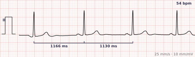
ECG criteria
| Feature | What you see in Sinus Bradycardia |
|---|---|
| Rate | Under 60 bpm. Below 40 is unusual and should prompt a search for block or drug effect. |
| Rhythm | Regular. Marked variation suggests sinus arrhythmia or sinus pauses. |
| P wave | Normal sinus P before every QRS — upright in II, inverted in aVR. |
| PR interval | 120–200 ms and constant. A long PR alongside bradycardia suggests first-degree AV block as well. |
| QRS | Narrow, under 120 ms. |
| ST-T | Usually normal. A prominent U wave becomes easier to see at slow rates. |
How to spot it
- Confirm the rate is under 60 by counting complexes across the strip, not by eye.
- Check every QRS still has a sinus P in front of it, with a constant PR. If some P waves are not conducted, this is AV block, not simple bradycardia.
- Look for pauses. A rhythm that is slow because of dropped beats is a different problem from one that is uniformly slow.
- Measure the PR — bradycardia plus a long PR often means the same cause is slowing both the node and conduction.
- Check the QRS is narrow. A slow wide-complex rhythm without P waves is an escape rhythm, not sinus bradycardia.
What it gets confused with
| Looks like | How to tell them apart |
|---|---|
| Junctional escape rhythm | Rate 40–60, narrow QRS, but no upright preceding P wave — P waves are absent, inverted or after the QRS. |
| Complete heart block | P waves and QRS complexes both present but unrelated — march them out separately and the PR varies randomly. |
| Sinus pause or arrest | Baseline sinus rate is normal, punctuated by dropped beats, rather than a uniformly slow rate. |
| 2:1 AV block | Twice as many P waves as QRS complexes. Look carefully at the T waves for a buried non-conducted P. |
| Atrial fibrillation with slow response | Irregularly irregular and no P waves. |
Traps
- Always count the P waves as well as the QRS complexes. A 2:1 block at an atrial rate of 80 gives a ventricular rate of 40 that looks like sinus bradycardia until you find the hidden P in each T wave.
- Sinus bradycardia at rest is normal in trained athletes and during sleep, where rates in the 40s are common and mean nothing.
- Beta blockers, calcium channel blockers, digoxin and amiodarone all cause it. Check the drug chart before investigating further.
- Bradycardia with an inferior STEMI is common and usually vagally mediated, but it can also be the first sign of AV nodal ischaemia.
Why it happens
The sinus node fires more slowly, most often because of increased vagal tone, drugs that suppress nodal automaticity, or intrinsic degeneration of the node itself. Conduction below the node is unaffected, which is why the P waves and QRS complexes still look normal.
Why it matters
Whether sinus bradycardia matters depends almost entirely on whether the patient has symptoms. An asymptomatic athlete at 45 needs nothing. A patient who is dizzy, syncopal or hypotensive at the same rate needs attention, because the slow rate is failing to deliver an adequate cardiac output.
Questions
Is sinus bradycardia normal in athletes?
Yes. Endurance training raises resting vagal tone, and resting rates in the 40s are common and asymptomatic in trained athletes. The rhythm is otherwise entirely normal sinus rhythm.
When does sinus bradycardia need treatment?
When it produces symptoms — dizziness, syncope, breathlessness or hypotension — or when it is the result of something correctable such as a drug. The rate alone does not decide it.
How do I tell sinus bradycardia from complete heart block?
In sinus bradycardia each QRS follows a P wave at a constant PR interval. In complete heart block the P waves and QRS complexes are independent, so the PR interval varies randomly and there are usually more P waves than QRS complexes.
Reading about sinus bradycardia is not the same as calling it on a tracing you have never seen.
Practise on real cases in ECG Pro