U Wave on the ECG
The U wave is a small deflection that follows the T wave on the ECG, normally under 25 per cent of the T wave amplitude and best seen in leads V2 and V3 at slow heart rates, becoming prominent in hypokalaemia and with certain drugs.
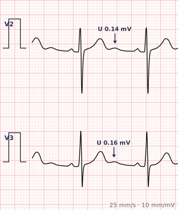
ECG criteria
| Feature | What you see in U Wave |
|---|---|
| Normal | Small, upright, under 25% of the T wave height. Most visible in V2–V3 and at slow rates. |
| Prominent | Over 25% of the T wave, or over 1–2 mm. Classically hypokalaemia. |
| Inverted | Abnormal in leads where the T is upright. Associated with ischaemia and hypertension. |
| Best leads | V2 and V3, where U waves are most reliably seen. |
| Rate | More visible at slow rates; merges with the T wave as rate increases. |
| Effect on QT | Merging with the T wave causes over-measurement of the QT interval. |
How to spot it
- Look just after the T wave in V2 and V3, where U waves show best.
- Compare the U wave height with the T wave in the same lead. Under a quarter is normal.
- If the U wave is prominent, check potassium and magnesium — hypokalaemia is the classic cause.
- Check whether the U wave is inverted in leads where the T is upright, which is abnormal.
- When measuring the QT, identify where the T ends and the U begins, and stop at the T.
- Look for the company hypokalaemia keeps: flattened T waves, ST depression and a long QU interval.
What it gets confused with
| Looks like | How to tell them apart |
|---|---|
| Hypokalaemia | Prominent U waves with flattened T waves and ST depression. The classic association. |
| Normal variant | Small upright U waves at slow heart rates, particularly in athletes. |
| Long QT | A U wave merging with the T is frequently misread as a prolonged QT. Distinguishing them is the point. |
| Drug effect | Antiarrhythmics, particularly class Ia and III agents, produce prominent U waves. |
| Hypercalcaemia or hypomagnesaemia | Other electrolyte causes of an abnormal repolarisation pattern. |
| Ischaemia | U wave inversion may accompany ischaemia and hypertension. |
Traps
- The single most important thing about the U wave is that it corrupts QT measurement. A U wave merging with the T produces a falsely prolonged QT and can lead to unnecessary alarm or the wrong drug being stopped.
- Prominent U waves should prompt a potassium measurement before anything else.
- Small U waves are entirely normal, particularly at slow heart rates and in athletes.
- U wave inversion in leads with upright T waves is abnormal and may accompany ischaemia, though it is a subtle and often overlooked sign.
- In severe hypokalaemia the U wave can become taller than the T wave, and the two together are sometimes misdescribed as a single bizarre T wave.
Why it happens
The origin of the U wave remains debated. The leading explanations are delayed repolarisation of the Purkinje fibres, late repolarisation of mid-myocardial M cells, or a mechanical after-potential generated by ventricular relaxation. Whatever the source, it represents the tail end of the repolarisation process, which is why it becomes prominent when repolarisation is prolonged — as in hypokalaemia.
Why it matters
The U wave matters in two ways. Prominent U waves are a recognisable and useful sign of hypokalaemia, which is common and correctable. And because the U wave sits immediately after the T, it is the most frequent cause of QT mismeasurement — making it a small waveform with a disproportionate influence on how the rest of the tracing is read.
Questions
What is a U wave on an ECG?
A small deflection following the T wave, normally less than a quarter of the T wave amplitude and best seen in leads V2 and V3, particularly at slow heart rates.
What causes prominent U waves?
Hypokalaemia is the classic cause. Prominent U waves also occur with hypomagnesaemia, bradycardia, and drugs that prolong repolarisation, particularly class Ia and class III antiarrhythmics.
Why does the U wave matter when measuring the QT interval?
Because a U wave merging with the T wave is easily included in the measurement, producing a falsely prolonged QT. Using the tangent method and stopping at the end of the T wave avoids this.
Are U waves always abnormal?
No. Small upright U waves are a normal finding, particularly at slow heart rates and in athletes. They become significant when prominent relative to the T wave, or when inverted in leads where the T is upright.
References
Reading about u wave is not the same as calling it on a tracing you have never seen.
Practise on real cases in ECG Pro