T Wave Inversion on the ECG
T wave inversion is a negative T wave in a lead where it should be upright, which is normal in aVR and often in V1 and lead III, but elsewhere may indicate ischaemia, strain, or a range of cardiac and non-cardiac conditions.
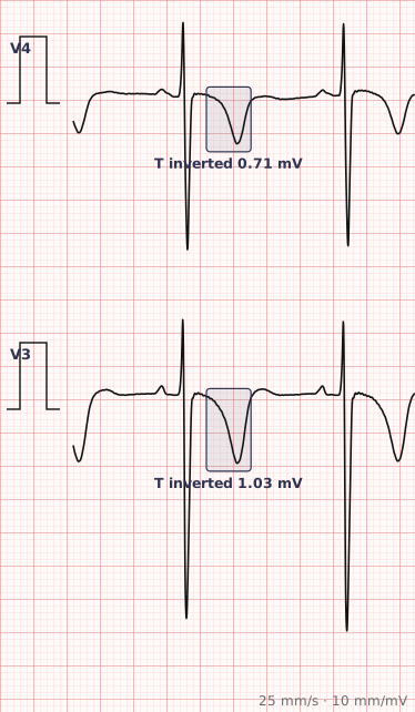
ECG criteria
| Feature | What you see in T Wave Inversion |
|---|---|
| Normal inversion | aVR always. V1 commonly. Lead III often. V1–V3 in children and some young adults (persistent juvenile pattern). |
| Abnormal | Inversion in two or more contiguous leads where the T should be upright. |
| Ischaemic | Deep, symmetric, narrow-based. Symmetry is the key feature. |
| Strain | Asymmetric — slow downstroke, rapid return — in leads with tall R waves, with downsloping ST depression. |
| Wellens pattern | Deep symmetric or biphasic T inversion in V2–V3 in a pain-free patient. Signals critical LAD stenosis. |
| Depth | Over 5 mm is a deep inversion; over 10 mm raises cerebral or apical causes. |
How to spot it
- First establish which leads normally have inverted T waves — aVR always, V1 usually, III often. Inversion there is not a finding.
- Identify inversion in two or more contiguous leads elsewhere, and name the territory.
- Assess symmetry. Symmetric inversion suggests ischaemia; asymmetric suggests strain or a secondary change.
- Check the R wave height in those leads. Tall R waves with asymmetric inversion points to hypertrophy.
- Check the QRS width. Bundle branch block produces expected discordant T inversion that should not be reported as ischaemia.
- Look specifically at V2 and V3 in anyone with recent chest pain — the Wellens pattern appears when the pain has settled and is easy to dismiss.
- Compare with a previous ECG. New inversion carries far more weight than established inversion.
What it gets confused with
| Looks like | How to tell them apart |
|---|---|
| Normal variant | aVR, V1 and III. Also V1–V3 in children and some young adults. |
| Ischaemia | Deep, symmetric, in a coronary territory, and new compared with previous tracings. |
| Wellens syndrome | Biphasic or deep symmetric inversion in V2–V3 with preserved R waves, in a pain-free patient. Critical LAD stenosis. |
| LVH strain | Asymmetric inversion in I, aVL, V5–V6 with tall R waves and downsloping ST depression. |
| Bundle branch block | Discordant inversion expected as a secondary change. |
| Pulmonary embolism | Inversion in V1–V4 and the inferior leads, with right heart strain. |
| Raised intracranial pressure | Deep, widespread, very broad T inversion with a long QT — cerebral T waves. |
Traps
- Wellens syndrome is the pattern not to miss. Biphasic or deep symmetric T inversion in V2 and V3, with preserved R waves, in a patient whose chest pain has resolved, indicates critical proximal LAD stenosis. The patient looks well and the ECG looks unremarkable at a glance.
- Know which leads are normally inverted before reporting anything. Inversion in aVR, V1 and III is expected.
- The persistent juvenile pattern — T inversion in V1 to V3 — is a normal variant in some young adults, particularly women.
- Symmetry is the most useful single feature. Ischaemic inversion is symmetric; strain inversion is not.
- Do not read T waves conventionally in bundle branch block, pre-excitation or a paced rhythm, where discordant inversion is expected.
Why it happens
The T wave records ventricular repolarisation, which normally proceeds from epicardium to endocardium — the opposite direction to depolarisation — producing an upright T wave concordant with the QRS. Anything that alters the sequence or timing of recovery inverts it: ischaemia delaying repolarisation in the affected region, hypertrophy changing recovery across a thickened wall, or abnormal depolarisation in bundle branch block forcing an abnormal recovery path.
Why it matters
T wave inversion covers an enormous range, from entirely normal to critical coronary stenosis, so its interpretation depends almost entirely on distribution, morphology and comparison with previous tracings. The Wellens pattern in particular is a case where an unimpressive-looking ECG in a comfortable patient identifies a lesion that warrants urgent angiography.
Questions
Which leads normally have inverted T waves?
Lead aVR always has an inverted T wave. Lead V1 commonly does, and lead III often does. Inversion in V1 to V3 is normal in children and persists into adulthood in some people as the persistent juvenile pattern.
What is Wellens syndrome?
A pattern of deeply inverted or biphasic T waves in leads V2 and V3, with preserved R wave progression, occurring in a pain-free patient after an episode of chest pain. It indicates critical stenosis of the proximal left anterior descending artery and carries a high risk of anterior infarction.
How do I tell ischaemic T inversion from a strain pattern?
Symmetry. Ischaemic T inversion is deep and symmetric with a narrow base. Strain inversion is asymmetric, with a gradual downstroke and rapid return, and occurs in leads with tall R waves alongside downsloping ST depression.
Are cerebral T waves a cardiac problem?
No. Very deep, broad, widespread T wave inversion with a prolonged QT interval can occur with raised intracranial pressure, particularly subarachnoid haemorrhage. The heart is usually structurally normal.
References
Reading about t wave inversion is not the same as calling it on a tracing you have never seen.
Practise on real cases in ECG Pro