Left Ventricular Hypertrophy (LVH) on the ECG
Left ventricular hypertrophy on the ECG is a pattern of increased QRS voltage in the leads facing the left ventricle, most often assessed by the Sokolow-Lyon or Cornell criteria, frequently accompanied by repolarisation abnormalities described as a strain pattern.
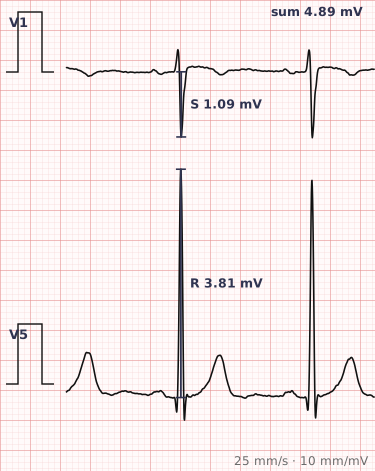
ECG criteria
| Feature | What you see in LVH |
|---|---|
| Sokolow-Lyon | S in V1 + R in V5 or V6 ≥ 35 mm. The most widely used and easiest to apply. |
| Cornell | R in aVL + S in V3 > 28 mm in men, > 20 mm in women. More specific than Sokolow-Lyon. |
| R in aVL | ≥ 11 mm alone is suggestive. |
| Strain pattern | Downsloping ST depression with asymmetric T inversion in I, aVL, V5–V6 — discordant to the tall R waves. |
| Axis | Often left. Marked left axis suggests coexisting fascicular block. |
| Other | Left atrial enlargement frequently accompanies it; QRS may be mildly prolonged. |
How to spot it
- Check the calibration first. A tracing recorded at half standard gain halves all voltages and hides LVH; double standard doubles them and invents it.
- Measure S in V1 and R in V5 or V6, and add them. 35 mm or more meets the Sokolow-Lyon criterion.
- Measure R in aVL and S in V3 for the Cornell criterion, which performs better in most populations.
- Look at the lateral leads for strain — downsloping ST depression with asymmetric T inversion, in the opposite direction to the tall R waves.
- Look for left atrial enlargement in lead II and V1, which commonly accompanies LVH and supports it.
- Check for confounders: bundle branch block, pre-excitation and a paced rhythm all make voltage criteria unreliable.
What it gets confused with
| Looks like | How to tell them apart |
|---|---|
| Thin chest wall or young age | High voltages with no strain pattern and no atrial enlargement. Common in slim young people and a frequent false positive. |
| Left bundle branch block | QRS 120 ms or more with a broad R in V6. Voltage criteria cannot be applied. |
| Lateral ischaemia | ST depression and T inversion without the tall R waves, and often with a different distribution. |
| Hypertrophic cardiomyopathy | Very high voltages with deep narrow Q waves in the lateral and inferior leads. |
| WPW | Short PR with a delta wave; the widened QRS can produce spurious voltage. |
Traps
- Always check the calibration mark before applying any voltage criterion. A 5 mm calibration pulse means half gain, and every measurement must be doubled.
- ECG voltage criteria are specific but insensitive. A normal ECG does not exclude left ventricular hypertrophy — echocardiography is the reference standard.
- Voltage alone in a thin young person is common and usually means nothing. The strain pattern and atrial enlargement are what add weight.
- Do not apply LVH criteria in the presence of left bundle branch block, pre-excitation or ventricular pacing. The activation sequence is already abnormal.
- Strain is easily reported as ischaemia. In LVH strain the ST depression is downsloping and the T inversion asymmetric, in leads with tall R waves.
Why it happens
A thickened left ventricular wall contains more myocardium, and a larger mass of tissue depolarising generates a larger electrical vector, which is recorded as increased voltage in the leads facing it. The strain pattern reflects altered repolarisation in the hypertrophied wall, where subendocardial perfusion is relatively reduced and the sequence of recovery is disturbed.
Why it matters
ECG evidence of left ventricular hypertrophy is an independent marker of cardiovascular risk, and its presence in hypertension changes how aggressively that hypertension is treated. On a practical level, LVH also distorts the rest of the tracing — it alters R wave progression, shifts the axis, and produces repolarisation changes that must not be confused with ischaemia.
Questions
What are the Sokolow-Lyon criteria for LVH?
The S wave in V1 added to the R wave in V5 or V6, whichever is taller, totalling 35 millimetres or more. It is the most widely used voltage criterion, though its sensitivity is limited.
What is a strain pattern?
Downsloping ST depression with asymmetric T wave inversion in the leads showing tall R waves, typically I, aVL, V5 and V6. It reflects abnormal repolarisation in hypertrophied myocardium rather than acute ischaemia.
Can LVH be diagnosed on an ECG alone?
The ECG can suggest it, but voltage criteria are specific rather than sensitive — many people with genuine hypertrophy have a normal ECG. Echocardiography is the reference standard for measuring wall thickness and mass.
Why does calibration matter so much for LVH?
Every voltage criterion is a measurement in millimetres, which depends entirely on the gain setting. At half standard gain all deflections are halved, so genuine hypertrophy is missed; at double gain, normal voltages appear pathological.
References
Reading about left ventricular hypertrophy is not the same as calling it on a tracing you have never seen.
Practise on real cases in ECG Pro