Myocardial Infarction (MI) on the ECG
Myocardial infarction on the ECG is a sequence of changes reflecting myocardial injury and necrosis, classically progressing from hyperacute T waves through ST elevation to T wave inversion and pathological Q waves, with the affected leads indicating the territory involved.
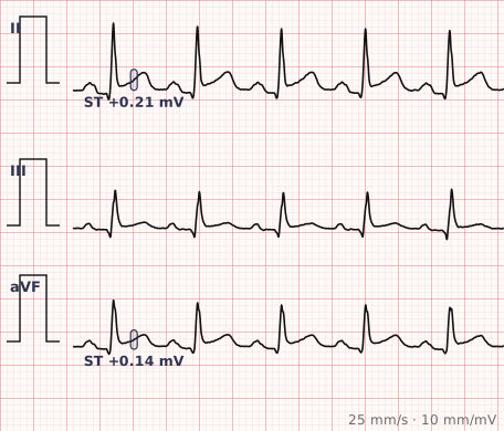
ECG criteria
| Feature | What you see in MI |
|---|---|
| Hyperacute T | Tall, broad, symmetric T waves. The earliest change, often within minutes and easily missed. |
| ST elevation | ≥ 1 mm in two contiguous leads — except V2–V3, where the cut-point is age and sex specific: ≥ 2.5 mm in men under 40, ≥ 2 mm in men 40 and over, ≥ 1.5 mm in women. |
| Reciprocal change | ST depression in the opposite territory. Its presence strongly supports true infarction over a mimic. |
| Q waves | Pathological when > 40 ms wide or > 25% of the R wave height. Develop over hours to days and usually persist. |
| T inversion | Follows the ST elevation as it resolves, over hours to days. |
| Territory | Inferior II, III, aVF · Anterior V1–V4 · Lateral I, aVL, V5–V6 · Posterior reciprocal changes in V1–V3. |
How to spot it
- Work through the leads in territory groups rather than in the order they are printed: inferior, anterior, lateral. Changes must be in two contiguous leads to count.
- Measure ST elevation at the J point, comparing against the TP segment as baseline.
- Look deliberately for reciprocal ST depression in the opposite territory. It is the most useful single discriminator from the mimics.
- Check for pathological Q waves — over 40 ms wide, or deeper than a quarter of the following R wave.
- Name the territory and therefore the likely vessel: inferior suggests the right coronary, anterior the left anterior descending, lateral the circumflex.
- If there is ST depression in V1–V3 with tall R waves, record posterior leads V7–V9 before concluding.
- In the presence of left bundle branch block or ventricular pacing, apply the Sgarbossa criteria rather than reading ST segments directly.
What it gets confused with
| Looks like | How to tell them apart |
|---|---|
| Pericarditis | Widespread concave ST elevation across territories, PR depression, and no reciprocal change. |
| Early repolarization | Concave ST elevation with notching at the J point, in a young person, stable over time. |
| Left ventricular hypertrophy | ST elevation in V1–V3 secondary to deep S waves, with voltage criteria met. |
| Left bundle branch block | Discordant ST elevation is expected. Use the Sgarbossa criteria. |
| Brugada syndrome | Coved ST elevation in V1–V2 with a partial RBBB appearance. |
| Takotsubo cardiomyopathy | Anterior ST elevation with deep T inversion and a long QT, and unobstructed coronaries. |
| WPW | Negative delta waves mimic pathological Q waves. Check the PR interval. |
Traps
- Reciprocal change is the most useful discriminator you have. Widespread ST elevation without reciprocal depression is much more likely to be pericarditis; elevation with clear reciprocal depression strongly favours infarction.
- Posterior infarction is the one most often missed. ST depression in V1 to V3 with tall R waves is the mirror image of posterior ST elevation — record posterior leads.
- A single ECG is a snapshot. The changes evolve, so repeat the tracing in a patient with ongoing symptoms and a normal first ECG.
- Right ventricular infarction accompanies up to a third of inferior infarcts. In inferior changes, record V4R before giving nitrates.
- Hyperacute T waves are the earliest sign and are routinely dismissed as normal. Tall, broad and symmetric T waves in a patient with chest pain deserve a repeat ECG.
- De Winter T waves — upsloping ST depression with tall symmetric T waves in the anterior leads — indicate proximal LAD occlusion without ST elevation.
Why it happens
Occlusion of a coronary artery deprives the myocardium it supplies of oxygen. Injured but still viable cells cannot maintain their resting membrane potential, generating a current of injury that displaces the ST segment towards the affected region — recorded as elevation in leads facing it and depression in leads facing away. When cells die, they no longer depolarise at all, leaving an electrical window through which the opposite wall is seen: the pathological Q wave.
Why it matters
ST elevation myocardial infarction is a time-critical diagnosis in which the ECG, not the troponin, drives the decision to reperfuse. Recognising the territory identifies the likely culprit vessel and predicts the complications to expect. Equally important is recognising the mimics, since inappropriate reperfusion carries real harm.
Questions
How much ST elevation is needed to diagnose a STEMI?
At least 1 millimetre in two contiguous leads, measured at the J point — in every lead except V2 and V3. Those two carry higher, age- and sex-specific cut-points: 2.5 millimetres in men under 40, 2 millimetres in men aged 40 and over, and 1.5 millimetres in women of any age. The leads must be contiguous and represent the same territory.
What is reciprocal change and why does it matter?
ST depression in leads facing away from the infarcted territory, produced by the same injury current viewed from the opposite direction. Its presence strongly supports true infarction and helps distinguish it from pericarditis, which typically causes widespread elevation without reciprocal depression.
Which leads show which territory?
Leads II, III and aVF face the inferior wall, usually the right coronary artery. Leads V1 to V4 face the anterior wall and the left anterior descending. Leads I, aVL, V5 and V6 face the lateral wall and the circumflex. Posterior infarction shows as reciprocal ST depression in V1 to V3.
How do I diagnose infarction in left bundle branch block?
The block already alters the ST segment, so the Sgarbossa criteria are used: concordant ST elevation of at least 1 millimetre, concordant ST depression of at least 1 millimetre in V1 to V3, or discordant ST elevation that is excessive relative to the QRS depth.
What makes a Q wave pathological?
Width greater than 40 milliseconds, or depth greater than 25 per cent of the height of the following R wave. Small narrow Q waves in the lateral leads are normal septal Q waves and are not pathological.
References
- Thygesen K et al. Fourth Universal Definition of Myocardial Infarction (2018). J Am Coll Cardiol.
- Rautaharju PM et al. AHA/ACCF/HRS Recommendations for the Standardization and Interpretation of the Electrocardiogram, Part IV: ST Segment, T and U Waves, and the QT Interval. Circulation 2009.
Reading about myocardial infarction is not the same as calling it on a tracing you have never seen.
Practise on real cases in ECG Pro