Early Repolarization on the ECG
Early repolarization is a common ECG pattern of J point elevation with notching or slurring at the end of the QRS complex, usually accompanied by concave ST elevation and prominent T waves, most often seen in young healthy people.
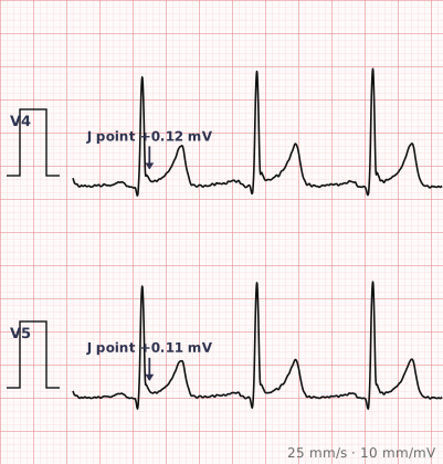
ECG criteria
| Feature | What you see in Early Repolarization |
|---|---|
| J point | Elevated with notching or slurring — the classic fish-hook appearance at the QRS-ST junction. |
| ST segment | Concave upward elevation, usually 1–4 mm. |
| Distribution | Most often V2–V5 and the inferior leads. Widespread rather than confined to a coronary territory. |
| T waves | Tall, prominent and concordant with the ST elevation. |
| Reciprocal change | Absent, other than the usual reciprocal appearance in aVR. |
| Stability | Unchanged across tracings over years. The most reassuring feature of all. |
How to spot it
- Note ST elevation, then look carefully at the J point for notching or slurring.
- Assess the ST shape. Concave upward fits early repolarization; convex or straightened should worry you.
- Map the distribution. Widespread across territories rather than confined to one is reassuring.
- Look for reciprocal ST depression. Its absence supports a benign cause.
- Consider the patient — young, healthy, often male, often athletic, and typically asymptomatic.
- Compare with any previous ECG. A pattern that is unchanged over years is the strongest evidence of all.
What it gets confused with
| Looks like | How to tell them apart |
|---|---|
| STEMI | Territorial, convex, with reciprocal depression, evolving over serial tracings. |
| Pericarditis | Widespread concave elevation with PR depression and PR elevation in aVR. |
| Left ventricular hypertrophy | Voltage criteria met, with a lateral strain pattern. |
| Brugada pattern | Coved elevation confined to V1–V2 with a partial RBBB appearance. |
| Hyperkalaemia | Peaked T waves with a widening QRS and flattened P waves. |
Traps
- Early repolarization is a diagnosis of exclusion in anyone with chest pain. The pattern is common enough that a patient with genuine infarction may also have it, so the clinical picture and serial ECGs matter more than the morphology.
- The single most useful investigation is an old ECG. An identical pattern years earlier effectively settles the question.
- Early repolarization in the inferior leads, particularly with a horizontal or downsloping ST segment after the J point, has been associated with a small increase in arrhythmic risk — the so-called J wave syndromes. The overwhelming majority of cases remain benign.
- It is more common in young men, in athletes, and in people of African descent, which is worth knowing so it is not over-investigated in those groups.
- The pattern may become less prominent with age and with increases in heart rate.
Why it happens
A transmural voltage gradient develops at the end of depolarisation, because repolarisation begins earlier in the epicardium than the endocardium. That gradient produces the J point elevation and the notch or slur at the junction of QRS and ST segment. The same transient outward potassium current that creates it is the one implicated, in rare cases, in the arrhythmic J wave syndromes.
Why it matters
Early repolarization is present in a substantial minority of healthy young adults and is overwhelmingly benign. Its practical importance is that it is a leading cause of unnecessary concern about ST elevation — recognising it, and confirming stability against an old tracing, prevents a great deal of over-investigation.
Questions
What is early repolarization on an ECG?
A pattern of J point elevation with notching or slurring at the end of the QRS complex, usually with concave ST elevation and prominent T waves, most often seen in the mid-precordial and inferior leads of young healthy people.
How do I distinguish early repolarization from a STEMI?
Early repolarization is concave, widespread rather than territorial, has no reciprocal ST depression, shows J point notching, and is unchanged across tracings. A STEMI is usually convex, confined to a territory, has reciprocal depression, and evolves over time.
Is early repolarization dangerous?
In the great majority of people it is entirely benign. Certain patterns, particularly inferior J waves with a horizontal or descending ST segment, have been associated with a small increase in arrhythmic risk, but the absolute risk remains very low.
Who most commonly has early repolarization?
It is most often seen in young adults, more frequently in men, in athletes, and in people of African descent. It tends to become less prominent with age.
Reading about early repolarization is not the same as calling it on a tracing you have never seen.
Practise on real cases in ECG Pro