Premature Atrial Contraction (PAC) on the ECG
A premature atrial contraction is an early heartbeat originating from an ectopic focus in the atria, producing a P wave of abnormal shape that arrives sooner than expected and is usually followed by a normal narrow QRS complex.
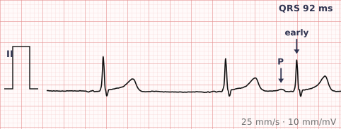
ECG criteria
| Feature | What you see in PAC |
|---|---|
| Timing | Early. The R-R interval before the beat is shorter than the underlying rhythm. |
| P wave | Present but abnormally shaped — different axis or morphology from the sinus P. Often hidden in the preceding T wave, deforming it. |
| PR interval | May differ from the sinus PR. A focus low in the atrium gives a shorter PR. |
| QRS | Normal and narrow — the impulse still uses the AV node and His-Purkinje system. Wide if conducted aberrantly. |
| Pause | Incomplete compensatory pause. The PAC resets the sinus node, so the pause is shorter than two full cycles. |
| ST-T | Normal, apart from the distortion where a hidden P sits on a T wave. |
How to spot it
- Find the beat that arrives early. March out the underlying rhythm first so you can see what "on time" would have looked like.
- Look for a P wave in front of it — that is what makes it atrial rather than ventricular.
- Compare that P wave with the sinus P waves. A different shape confirms a different origin.
- Check the preceding T wave. A T that is taller, notched or peaked compared with the others usually has the ectopic P buried in it.
- Measure the pause. Mark two sinus cycles from the beat before the PAC — if the next sinus beat comes early relative to that mark, the pause is incomplete, which fits an atrial origin.
What it gets confused with
| Looks like | How to tell them apart |
|---|---|
| Premature ventricular contraction | PVCs are wide with no preceding P wave and a full compensatory pause. PACs are narrow with an abnormal P. |
| Sinus arrhythmia | Rate varies gradually with respiration; every P wave is identical in shape. |
| Blocked PAC | An early P wave with no QRS after it, producing a pause. Commonly misread as sinus arrest or AV block. |
| PAC with aberrancy | Early, wide, but with a preceding abnormal P wave — the P is what separates it from a PVC. |
| Wandering atrial pacemaker | A continuously shifting pacemaker with three or more P shapes, not isolated early beats on a regular background. |
Traps
- A blocked PAC is the most common cause of an unexpected pause, and is regularly misdiagnosed as sinus arrest or second-degree AV block. Always look for a hidden early P wave deforming the T that precedes the pause.
- The compensatory pause distinction is genuinely useful: PACs reset the sinus node so the pause is incomplete, PVCs usually do not so the pause is full.
- PACs conducted with aberrancy look wide and get called PVCs. The preceding abnormal P wave settles it.
- Occasional PACs are found in most healthy people and mean nothing on their own. Frequent PACs, though, raise the likelihood of later atrial fibrillation.
Why it happens
An atrial focus outside the sinus node depolarises before the next sinus impulse is due, and captures the atria from that site. Because the impulse then reaches the AV node and travels down the normal conduction system, the QRS is unchanged. The ectopic impulse also travels back into the sinus node and resets its timing, which is why the following pause is incomplete.
Why it matters
Isolated PACs are common, benign and need no treatment. Their significance is mostly as a mimic — of pauses, of AV block, and of ventricular ectopy — and as a marker: a high burden of atrial ectopy is associated with an increased risk of developing atrial fibrillation.
Questions
How do I tell a PAC from a PVC?
Look for a P wave and measure the QRS. A PAC has an abnormal P wave in front of a narrow QRS and an incomplete compensatory pause. A PVC has no preceding P wave, a wide bizarre QRS, and usually a full compensatory pause.
What is a blocked PAC?
An atrial ectopic that arrives so early the AV node is still refractory, so no QRS follows. It produces a pause with a hidden P wave sitting on the preceding T wave, and is frequently mistaken for sinus arrest or AV block.
Are premature atrial contractions dangerous?
Isolated PACs occur in most healthy people and are benign. A high burden is associated with an increased likelihood of developing atrial fibrillation, so frequent PACs are worth noting rather than ignoring.
Reading about premature atrial contraction is not the same as calling it on a tracing you have never seen.
Practise on real cases in ECG Pro