Premature Ventricular Contraction (PVC) on the ECG
A premature ventricular contraction is an early heartbeat originating from a focus within the ventricles, producing a wide and abnormally shaped QRS complex with no preceding P wave, usually followed by a full compensatory pause.
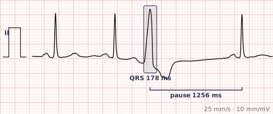
ECG criteria
| Feature | What you see in PVC |
|---|---|
| Timing | Early, arriving before the next sinus beat is due. |
| P wave | None before the QRS. A retrograde P may follow it. Sinus P waves continue underneath, undisturbed. |
| QRS | Wide (over 120 ms) and bizarre, unlike any conducted beat on the tracing. |
| ST-T | Discordant — the ST segment and T wave point opposite to the main QRS deflection. Concordant repolarisation suggests something else. |
| Pause | Full compensatory pause — the interval spanning the PVC equals two normal R-R intervals, because the sinus node is not reset. |
| Morphology | Unifocal PVCs all look the same. Multifocal PVCs have differing shapes, implying more than one focus. |
How to spot it
- Spot the beat that is both early and wide. That combination alone puts a PVC at the top of the list.
- Check there is no P wave immediately before it. A preceding abnormal P would make it an aberrantly conducted PAC instead.
- Confirm the ST-T is discordant. A wide beat whose T wave points the same way as the QRS should make you look again.
- Measure the pause: mark two normal R-R intervals from the beat before. If the next sinus beat lands exactly on the mark, the pause is fully compensatory.
- Describe the pattern and burden — isolated, bigeminy, couplets, unifocal or multifocal. Those descriptors carry more information than the count.
What it gets confused with
| Looks like | How to tell them apart |
|---|---|
| PAC with aberrant conduction | An abnormal P wave precedes the wide beat, and the pause is incomplete. |
| Ventricular tachycardia | Three or more consecutive ventricular beats above 100. A run of PVCs becomes VT by definition at that point. |
| Fusion beat | A hybrid shape, intermediate between the sinus QRS and the PVC, from simultaneous activation by both. |
| Paced beat | A pacing spike immediately precedes the wide complex. |
| Escape beat | Late, not early. Same wide morphology, but it arrives after a pause rather than interrupting the rhythm. |
Traps
- The single most useful distinction is early versus late. An early wide beat is a PVC. A late wide beat after a pause is an escape beat, and suppressing it can be catastrophic.
- R-on-T, where a PVC lands on the preceding T wave, can precipitate polymorphic VT. It matters most when the QT is long.
- Multifocal PVCs imply more than one irritable focus and generally carry more weight than the same number of unifocal beats.
- A very frequent PVC burden, above roughly 10 to 20% of beats, can itself cause a cardiomyopathy — one of the few situations where ectopy is treated for its own sake.
- PVCs are common in healthy hearts. Context decides significance: the same tracing means different things in a fit 25-year-old and in someone with a previous infarct.
Why it happens
A focus of ventricular myocardium depolarises spontaneously before the next sinus impulse arrives. Because it starts below the His-Purkinje system, activation spreads slowly from muscle cell to muscle cell rather than through fast conducting fibres, which produces the wide and unusually shaped QRS. Repolarisation follows the same abnormal path, giving the discordant ST-T.
Why it matters
PVCs are extremely common and usually benign in a structurally normal heart. They matter more when they are frequent, multifocal, occur in runs, or appear in the context of structural disease or a long QT interval. Recognising them accurately mostly prevents over-treatment — and prevents an escape beat being mistaken for one.
Questions
What is a compensatory pause and why does it follow a PVC?
The ventricular ectopic does not usually travel backwards into the sinus node, so the node keeps its own timing. The next sinus impulse arrives on schedule but finds the ventricle refractory, so it is not conducted, and the interval spanning the PVC equals exactly two normal cycles.
Are PVCs dangerous?
In a structurally normal heart, usually not. Concern rises with frequency, multiple morphologies, runs of three or more, a very high burden that can itself weaken the ventricle, and underlying structural heart disease.
What is R-on-T?
A premature ventricular beat that falls on the T wave of the preceding beat, during the vulnerable period of repolarisation. It can trigger polymorphic ventricular tachycardia, and is of particular concern when the QT interval is prolonged.
What is the difference between unifocal and multifocal PVCs?
Unifocal PVCs all share the same shape, implying a single origin. Multifocal PVCs vary in shape, implying several irritable foci, and generally carry more clinical weight.
Reading about premature ventricular contraction is not the same as calling it on a tracing you have never seen.
Practise on real cases in ECG Pro