T Wave Change on the ECG
T wave change is any abnormality of T wave shape, amplitude or direction that falls short of a specific diagnosis, including flattening, low amplitude, peaking, biphasic morphology and inversion in leads where the T wave should be upright.
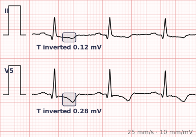
ECG criteria
| Feature | What you see in T Wave Change |
|---|---|
| Flattening | T wave amplitude under 10% of the R wave, or nearly isoelectric. Common and highly non-specific. |
| Peaking | Tall, narrow, symmetric T waves. Think hyperkalaemia first, and hyperacute ischaemia second. |
| Biphasic | Up-then-down or down-then-up. In V2–V3 with recent chest pain this is the Wellens pattern. |
| Inversion | Abnormal outside aVR, V1 and III. Symmetric favours ischaemia; asymmetric favours strain. |
| Normal variants | Inverted T in aVR always. V1 commonly. V1–V3 in children and the persistent juvenile pattern. |
| Distribution | Territorial grouping raises concern; scattered change is usually non-specific. |
How to spot it
- Establish which leads normally carry an inverted or low T wave, so those are not reported as abnormal.
- Characterise the change: flat, peaked, biphasic or inverted. The category directs the differential.
- Peaked T waves are the ones that need immediate action — check potassium, and look for QRS widening and loss of P waves.
- Assess symmetry. Symmetric change favours ischaemia; asymmetric favours a strain or secondary pattern.
- Look in V2 and V3 specifically after chest pain, for the biphasic Wellens pattern.
- Check the QRS. Bundle branch block, pre-excitation and pacing all produce expected T wave change.
- Compare with a previous tracing before deciding the change is new.
What it gets confused with
| Looks like | How to tell them apart |
|---|---|
| Hyperkalaemia | Tall peaked symmetric T waves, later with a widening QRS and flattened P waves. The emergency in this list. |
| Hyperacute ischaemia | Broad, tall, symmetric T waves in a coronary territory, in a patient with chest pain. Earliest sign of infarction. |
| Wellens syndrome | Biphasic or deep symmetric T inversion in V2–V3, pain-free, with preserved R waves. Critical LAD stenosis. |
| Hypokalaemia | Flattened T waves with prominent U waves and ST depression. |
| LVH strain | Asymmetric inversion in leads with tall R waves. |
| Persistent juvenile pattern | T inversion in V1–V3 in a young adult, unchanged over years. A normal variant. |
| Cerebral T waves | Very deep, broad, widespread inversion with a long QT, in raised intracranial pressure. |
Traps
- Peaked T waves deserve a potassium level before anything else. Hyperkalaemia is the one cause on this list that can kill within hours and is trivially correctable.
- T wave flattening is so common and so non-specific that it is close to meaningless in isolation — but it should not be used to dismiss a symptomatic patient either.
- Know the normal variants before reporting. Inversion in aVR, V1 and III is expected, and the persistent juvenile pattern is normal in some young adults.
- Hyperacute T waves are the earliest change of infarction and are routinely dismissed as normal because they are upright and tall rather than obviously abnormal.
- Do not read T waves conventionally in bundle branch block, pre-excitation or a paced rhythm, where discordant change is expected.
Why it happens
The T wave records ventricular repolarisation, and its shape depends on the gradient of recovery across the ventricular wall. That gradient is altered by ischaemia, by wall thickness, by the sequence of depolarisation, and directly by extracellular potassium, which changes the slope of the repolarising phase of the action potential — the reason hyperkalaemia produces such a characteristic tall, narrow, symmetric T wave.
Why it matters
Most T wave change is non-specific and of limited value on its own. Its importance lies in the small number of patterns that are not: peaked T waves signalling hyperkalaemia, hyperacute T waves as the first sign of coronary occlusion, and the Wellens pattern marking critical proximal stenosis. Recognising those three inside the noise is the whole skill.
Questions
What does non-specific T wave change mean?
That the T wave shape, amplitude or direction is abnormal but does not meet criteria for a specific diagnosis. It is common on routine ECGs and carries little weight in isolation.
What do peaked T waves mean?
Most importantly hyperkalaemia, which produces tall, narrow, symmetric T waves and can progress to QRS widening, loss of P waves and cardiac arrest. Broad, tall, symmetric T waves in a chest pain patient may instead be the hyperacute phase of infarction.
Which T wave inversions are normal?
Lead aVR always, lead V1 commonly, and lead III often. Inversion in V1 to V3 is normal in children and persists into adulthood in some people as the persistent juvenile pattern.
How do I tell ischaemic T wave change from a strain pattern?
Symmetry and company. Ischaemic change tends to be symmetric and territorial; strain change is asymmetric, occurs in leads with tall R waves, and accompanies downsloping ST depression and voltage criteria for hypertrophy.
References
Reading about t wave change is not the same as calling it on a tracing you have never seen.
Practise on real cases in ECG Pro