Supraventricular Tachycardia (SVT) on the ECG
Supraventricular tachycardia is a regular narrow-complex tachycardia arising above the ventricles, typically at 150 to 250 beats per minute, in which P waves are absent, buried in the QRS complex, or visible only as a distortion at its end.
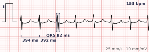
ECG criteria
| Feature | What you see in SVT |
|---|---|
| Rate | 150–250 bpm, most often around 180. Rigidly constant. |
| Rhythm | Regular, and metronomically so — beat-to-beat variation is essentially nil. |
| P wave | Not visible, or seen as a retrograde P distorting the terminal QRS — a pseudo-R' in V1 or a pseudo-S in the inferior leads. |
| PR interval | Not measurable. |
| QRS | Narrow, under 120 ms. Wide if there is rate-related aberrancy or pre-existing bundle branch block. |
| ST-T | ST depression is very common at these rates and usually rate-related rather than ischaemic. |
How to spot it
- Confirm the rate is over 150 and the rhythm is rigidly regular. Variation of even a few milliseconds between beats argues against SVT.
- Confirm the QRS is narrow. A wide regular tachycardia should be treated as ventricular tachycardia until proven otherwise.
- Hunt for atrial activity. Compare the QRS shape with an old tracing in sinus rhythm — a small extra deflection at the end of the QRS in SVT is a retrograde P wave, not part of the QRS.
- Rule out flutter by looking for a sawtooth baseline in II, III, aVF and V1. Flutter at 150 is the main trap.
- Note whether onset and offset are abrupt. SVT starts and stops like a switch; sinus tachycardia accelerates and decelerates.
What it gets confused with
| Looks like | How to tell them apart |
|---|---|
| Sinus tachycardia | Visible sinus P waves, a rate that drifts with activity, and gradual onset. SVT is fixed and starts abruptly. |
| Atrial flutter with 2:1 block | Sawtooth flutter waves and an atrial rate near 300. The mimic that catches people out most. |
| Ventricular tachycardia | Wide QRS. If a regular wide tachycardia is in front of you, assume VT — being wrong the other way is far more dangerous. |
| Atrial fibrillation with rapid response | Irregular. SVT is regular. |
Traps
- A regular wide-complex tachycardia is ventricular tachycardia until proven otherwise, even in a well-looking young patient. SVT with aberrancy is the less likely and less dangerous of the two possibilities.
- The ST depression seen at 200 bpm is usually demand-related and resolves on reversion. Do not commit to a diagnosis of ischaemia on the tachycardic tracing.
- Compare with an old ECG whenever one exists. A small terminal deflection that is present in tachycardia but absent in sinus rhythm is a retrograde P wave and confirms the diagnosis.
- Record a rhythm strip during adenosine or a vagal manoeuvre. The moment of termination is often the most diagnostic few seconds of the whole encounter, and it cannot be recovered afterwards.
Why it happens
Most cases are re-entrant. In AV nodal re-entrant tachycardia the circuit sits within the AV node itself, using fast and slow pathways; in AV re-entrant tachycardia it uses an accessory pathway between atrium and ventricle. Because the circuit conducts down the normal His-Purkinje system, the QRS stays narrow, and because the loop is fixed in size, the rate barely varies.
Why it matters
SVT is rarely life-threatening in a structurally normal heart, but it is frequently symptomatic and recurrent, and it is one of the few arrhythmias that can be cured outright by ablation. Its practical importance on the ECG is that it must be distinguished from ventricular tachycardia and from flutter, both of which are managed very differently.
Questions
What rate distinguishes SVT from sinus tachycardia?
There is no clean cut-off, but a resting adult above about 180 is far more likely to be in SVT. More useful than rate is the pattern: SVT is fixed and starts abruptly, whereas sinus tachycardia drifts up and down with the patient's state.
Where is the P wave in SVT?
Usually buried inside the QRS complex, because atria and ventricles depolarise almost simultaneously. When visible it is retrograde and falls at the end of the QRS, appearing as a pseudo-R' in V1 or a pseudo-S wave in the inferior leads.
Is a wide-complex tachycardia ever SVT?
Yes, when there is pre-existing bundle branch block or rate-related aberrant conduction. But ventricular tachycardia is both more common and more dangerous, so a regular wide-complex tachycardia should be managed as VT unless there is clear evidence otherwise.
Why does ST depression appear during SVT?
At very high rates, diastole shortens and coronary filling falls while myocardial demand rises. The resulting ST depression is usually rate-related and resolves once sinus rhythm returns.
Reading about supraventricular tachycardia is not the same as calling it on a tracing you have never seen.
Practise on real cases in ECG Pro