Left Anterior Fascicular Block (LAFB) on the ECG
Left anterior fascicular block is a conduction delay in the anterior fascicle of the left bundle branch, producing marked left axis deviation between −45 and −90 degrees with a qR pattern in aVL, an rS pattern in the inferior leads, and a QRS duration that remains normal or only slightly prolonged.
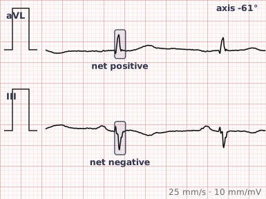
ECG criteria
| Feature | What you see in LAFB |
|---|---|
| Axis | −45° to −90° — marked left axis deviation. This is the defining feature. |
| QRS | Normal or only slightly prolonged, under 120 ms. Widening suggests something else as well. |
| I, aVL | qR pattern — small Q, tall R. |
| II, III, aVF | rS pattern — small R, deep S. S wave deeper in III than in II. |
| R peak time | Delayed in aVL, at 45 ms or more. |
| ST-T | Usually normal. |
How to spot it
- Check the axis first. Lead I positive with aVF negative means left axis deviation; work out whether it is beyond −45° by looking at lead II.
- Look at lead II. If lead II is predominantly negative, the axis is more negative than −30° and fascicular block becomes likely.
- Confirm the pattern: qR in I and aVL, rS in II, III and aVF.
- Measure the QRS width. Under 120 ms with marked left axis fits LAFB; a wide QRS points to LBBB or a non-specific delay instead.
- Exclude the mimics — inferior infarction and left ventricular hypertrophy both shift the axis leftward.
- Check for coexisting RBBB, which together make bifascicular block.
What it gets confused with
| Looks like | How to tell them apart |
|---|---|
| Inferior myocardial infarction | Pathological Q waves in II, III and aVF rather than small r waves. LAFB has an rS, not a QS. |
| Left ventricular hypertrophy | Voltage criteria met; axis shift is usually less extreme than −45°. |
| Left bundle branch block | QRS 120 ms or more with a broad R in V6. LAFB keeps a narrow QRS. |
| Left posterior fascicular block | Right axis deviation instead, with rS in I and qR in III. Much rarer. |
| Simple left axis deviation | Axis between 0 and −30°, without the fascicular block pattern. |
Traps
- Lead II is the quick test. A predominantly negative lead II means the axis is beyond −30°, which is the threshold that makes fascicular block worth considering.
- Inferior infarction is the important mimic. Look for genuine pathological Q waves in the inferior leads rather than the small initial r wave that LAFB preserves.
- LAFB can mask or mimic other findings: it may conceal inferior infarction, and it can produce poor R wave progression in the anterior leads.
- LAFB with RBBB is bifascicular block; adding first-degree AV block has traditionally been called trifascicular block and raises concern about progression.
- Isolated LAFB is common with age and hypertension, and by itself is of limited significance.
Why it happens
The left bundle branch divides into an anterior and a posterior fascicle. When the anterior fascicle fails to conduct, the left ventricle is activated first through the posterior fascicle — inferiorly and to the right — before the impulse spreads back up and leftward to the anterolateral wall. That reversed sequence swings the mean QRS vector sharply leftward and superiorly, producing the marked left axis deviation, while overall activation remains fast enough to keep the QRS narrow.
Why it matters
Left anterior fascicular block is common, particularly with increasing age, hypertension and coronary disease, and in isolation it carries little weight. It matters chiefly as a component of bifascicular block, as a mimic of inferior infarction, and as a cause of altered R wave progression that can lead to other findings being over-read.
Questions
How do I recognise left anterior fascicular block?
Marked left axis deviation between −45 and −90 degrees, a qR pattern in lead aVL, an rS pattern in the inferior leads, and a QRS duration that remains under 120 milliseconds.
Why does the QRS stay narrow in fascicular block?
Only one of the two left-sided fascicles is blocked, so the left ventricle is still activated through the other one and through the fast conducting system. Total activation time is barely prolonged, unlike complete bundle branch block.
How is LAFB distinguished from inferior myocardial infarction?
Both shift the axis leftward, but LAFB preserves a small initial r wave in the inferior leads, giving an rS pattern. Inferior infarction produces pathological Q waves or a QS complex instead.
What is bifascicular block?
Block of two of the three main conduction fascicles — most often right bundle branch block together with left anterior fascicular block. It reflects more extensive conduction disease than either finding alone.
References
Reading about left anterior fascicular block is not the same as calling it on a tracing you have never seen.
Practise on real cases in ECG Pro